Research: Histopathology of the atherosclerotic plaque and of the carcinogenic processes
Principal Investigator: Prof. Alessandro Mauriello

Pathophysiology of the vascular system
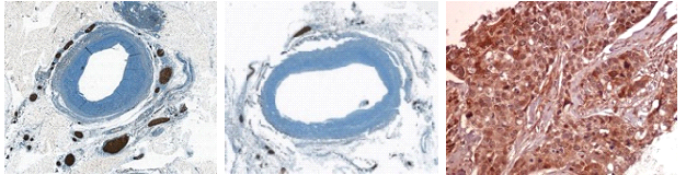
Clinical studies demonstrated that some patients are asymptomatic for life, even though they harbor atherosclerotic plaques in their vasculature, whereas others suffer ischemic symptoms such as myocardial infarction and stroke. The first condition is usually characterized by a slow growing of a silent “stable plaque”, whereas clinical events are associated to one or more “unstable plaques” .
Our Research group has paved the way for the understanding of the role of inflammation in acute coronary syndromes by demonstrating, using morphological studies, that the principal cause of coronary instability is not to be found in the vulnerability of a single atherosclerotic plaque, but in the presence of multiple vulnerable plaques in the entire coronary tree, associated with a widespread inflammatory process. Other researchs on the carotid plaques have helped to better clarify the pathogenesis of acute cerebrovascular syndromes first demonstrating by histology that plaque rupture and thrombosis are the principal determinants of major stroke, regardless of the degree of carotid stenosis. The carotid plaque remains up to 2 years at a stage of chronic destabilization, underlining the importance of early surgical intervention to reduce the risk of subsequent events. Moreover, it is possible to hypothesize that plaque stability is influenced not only by local, but also by systemic factors. To this end recently we have studied the contribution of the sympathetic nervous system to the development of hypertension. Several indirect evidences have found that the sympathetic overactivity in hypertensive patients is caused by neurogenic signals originating in the damaged kidneys. We performed an histo-morphometric evaluation of two and three dimensional distribution of renal nerves in hypertensive (H) vs. normotensive (N) individuals and demonstrated that renal nerves are increased in H patients as compared to N patients, providing the morphological basis for the renal denervation, that today represents the most important emerging treatment for resistant hypertension.
In situ identification of new diagnostic and prognostic bio-markers in tumors.
The characterization of molecular pathways that connect altered inflammatory response and cancer progression could provide the scientific rationale for the identification of new prognostic and predictive biomarkers. To date, the expression of programmed death ligand (PD-L1) allows cancer cells to escape the immune response of T lymphocytes, and thus, to trigger the metastatic process. In particular we investigated in the prostate tumors the correlation between the composition of intratumoral inflammatory infiltrate and the expression of PD-L1 by cancer cells. Our results highlighted a significant increase of the number of PD-L1 positive cells in prostate tumors respect to benign lesions, associated to a decrease of both PD1 positive lymphocytes and tumor-infiltrated macrophages, mainly M2 subpopulation. These data could be useful to develop therapeutical strategies able to inhibit the PD-L1 activity and, at the same time, to re-activate the antitumor inflammatory process. Ongoing research are evaluating the the key role of host immune-surveillance in the triple negative breast cancers, a class of invasive breast cancers which are refractory to the currently available therapies. In addition, on prostatic cancer we demonstrated the association between the presence of cancer cells with morphological and molecular characteristics of osteoblasts (prostate osteoblast-like cells—POLCs),and the development of bone metastasis. The POLCs’ origin was linked to epithelial to mesenchymal transformation (EMT) and subsequent osteoblastic differentiation stimulated by bone morphogenetics protein (BMP)-2.
The definition of the epigenetic profile and the corresponding protein expression in the biopsies of tumor tissues, comparing them with healthy tissues, is necessary not only for an early diagnosis, but also for identifying new target molecules therapeutically useful, in order to switch from a standardized therapy to personalized treatment that in the future could increase survival and improve the quality of life of patients. In collaboration with TOR research group we demonstrated, evaluating by immunohistochemistry the expression in human surgical biopsies, that ZNF185 is a p63 target gene critical for epidermal differentiation and squamous cell carcinoma (SCC) development. Therefore, ZNF185 could be considered a potential specific biomarker for SCC diagnosis and prognosis. Ongoing studies are focusing the expression of the various ZNF proteins in other human tumors with the aim to detect cellular changes which early occur in the neoplastic transformation.